DIAGNOSTIC IMAGING

Full range of portable digital equipment with
images stored online for immediate availability.

Contact Us

(08) 9296 6666 (office hours & after-hours emergency)

admin@belvoirequinehospital.com.au

Lot 158 West Swan Rd, Belhus, WA 6069

Ultrasonography

Ultrasonography uses sound energy at very high frequencies to produce visual images of internal soft tissue structures such as tendons, muscle, abdominal and reproductive organs.

At Belvoir Equine Hospital, we offer both internal and external ultrasonography. Internal ultrasonography is used mostly during the breeding season to evaluate the reproductive organs of mares and to assess and monitor foetal well-being. Scanning internally can also provide information on important abdominal structures such as the intestine, bladder, kidneys and spleen.

For more information on the reproductive services we provide, please – Click Here
A number of soft tissue structures can also be evaluated externally. Diagnostic ultrasound is used to examine and assess;

Tear that has caused a hole in the tendon.

Tendons: Tendinitis (‘bowed tendon’) is a common problem in horses. It is the result of tendon fibre disruption often due to excessive strain.

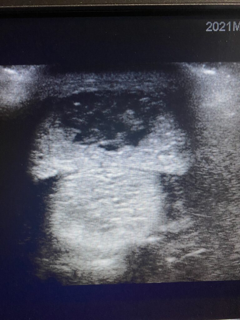
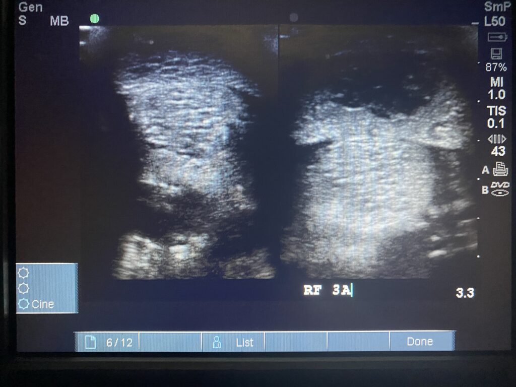
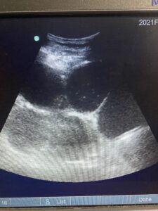
Abdomen:  Numerous organs can beassessed ultrasonographically, including the stomach, liver, intestines, spleen, kidneys and uterus. We routinely scan the abdomen in horses and foals with colic and suspected liver problems. It also facilitates evaluation of foetal well being in pregnant mares.

Joints:  Joint surfaces, synovial fluid, cartilage and supporting soft tissue structures can be evaluated. This is often used in foals with suspected ‘joint ill’ or young horses with suspected OCD.

Chest: This is particularly useful for evaluation and management of ‘rattles’ (Rhodococcus equi) and pleuropneumonia (for horses with travel sickness for example). The heart and its function can also be evaluated.

Loops of extended bowel

Radiography:

Radiography utilises x-rays to visualise the structure and integrity of bones. It is a very common diagnostic tool for lameness evaluation and assessment of bones secondary to trauma. Due to the size of horses, it is infrequently used to assess the chest or abdominal structures.

Currently at the clinic, we are using digital radiography which involves the image being developed in a digital format and displayed on a computer screen. Digital radiography has revolutionised the extent to which we can examine bone structure. The images are processed instantly and have excellent clarity and detail. **We can travel to you for radiographs in emergencies, where the horse cannot be moved.

Video Endoscopy:

Endoscopy is facilitated through the use of an endoscope, which is made up of optical fibres and a lens. It allows us to examine some internal structures of the head including; the nasal passages, sinus drainage angles, pharynx, guttural pouches and larynx. The ‘windpipe’ or trachea can also be evaluated and this is particularly useful for evaluating respiratory disease. We also use endoscopes to examine the oesophagus and stomach and therefore they are useful in cases of choke or gastric ulceration.

Here at Belvoir Equine Clinic, we currently have video endoscopy which projects the image onto a screen so what we see can be clearly explained to you. We routinely use endoscopy here at BEC for work-up of poor performance (or wind problems), respiratory disease and sinus problems.