Ultrasound Investigations
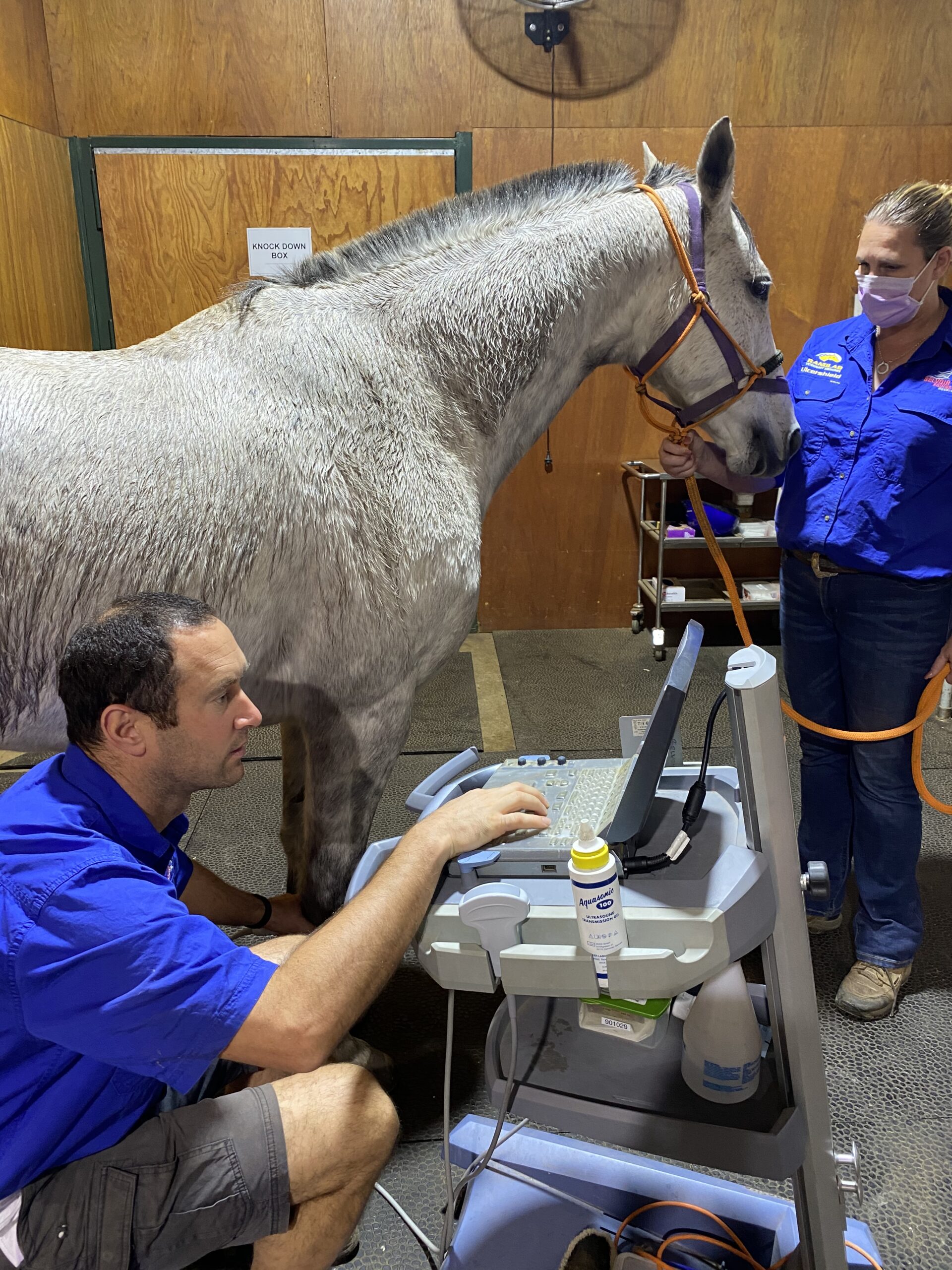
We offer both internal and external
ultrasonography.
Contact Us
(08) 9296 6666 (office hours & after-hours emergency)
admin@belvoirequinehospital.com.au
Lot 158 West Swan Rd, Belhus, WA 6069
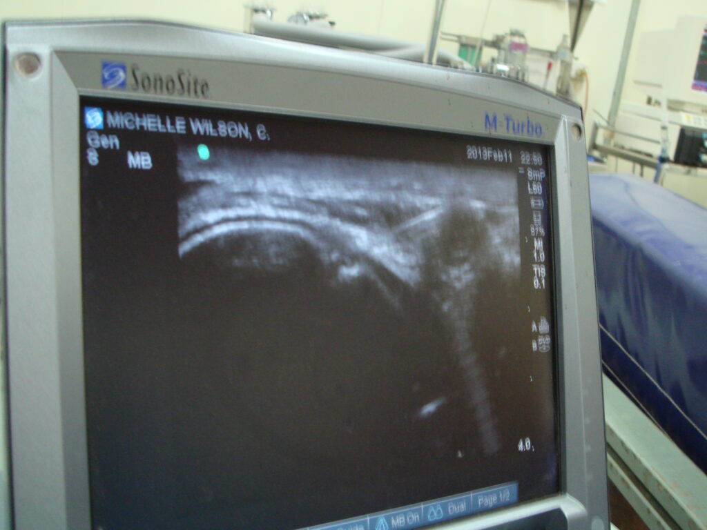
Our practice currently uses a state of the art dual frequency ultrasound machine to enhance our diagnostic and therapeutic capabilities.
These allow imaging of the majority of the equine body, including many areas that are not readily accessible to external examination.
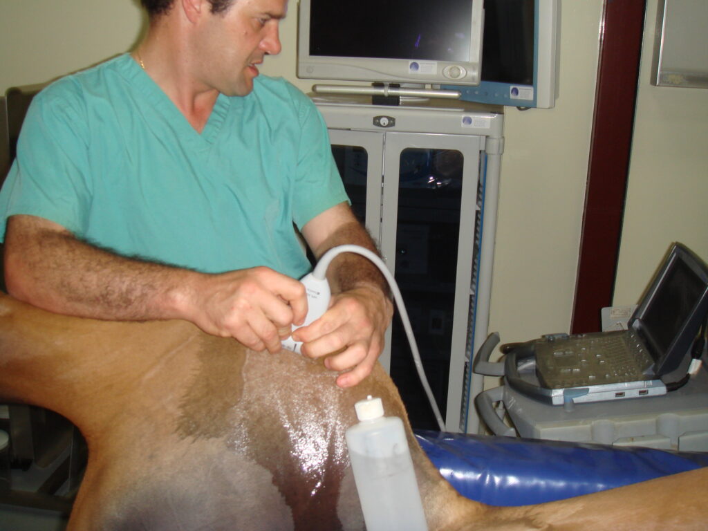
Ultrasonography:
Ultrasonography uses sound energy at very high frequencies to produce visual images of internal soft tissue structures such as tendons, muscle, abdominal and reproductive organs
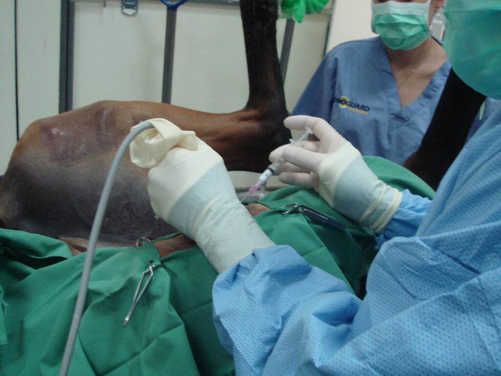
At Belvoir Equine Clinic, we offer both internal and external ultrasonography. Internal ultrasonography is used mostly during the breeding season to evaluate the reproductive organs of mares and to assess and monitor foetal well-being. Scanning internally can also provide information on important abdominal structures such as the intestine, bladder, kidneys and spleen.
For more information on the reproductive services we provide, please – Click Here
A number of soft tissue structures can also be evaluated externally. Diagnostic ultrasound is used to examine and assess;
Tendons: Tendinitis (‘bowed tendon’) is a common problem in horses. It is the result of tendon fibre disruption often due to excessive strain.
Joints: Joint surfaces, synovial fluid, cartilage and supporting soft tissue structures can be evaluated. This is often used in foals with suspected ‘joint ill’ or young horses with suspected OCD.
Abdomen: Numerous organs can be assessed ultrasonographically, including the stomach, liver, intestines, spleen, kidneys and uterus. We routinely scan the abdomen in horses and foals with colic and suspected liver problems. It also facilitates evaluation of foetal well being in pregnant mares.
Chest: This is particularly useful for evaluation and management of ‘rattles’ (Rhodococcus equi) and pleuropneumonia (for horses with travel sickness for example). The heart and its function can also be evaluated.